AP ABDOMEN UPRIGHT
Anteroposterior abdomen projection protocol in erect position
MAIN INDICATION AND PRIORITY
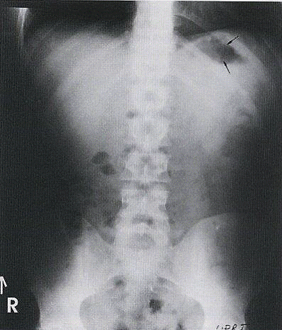
Pathologies demonstrated: Abnormal masses, air-fluid levels, and intraperitoneal air collections under diaphragm.
OBTAIN UPRIGHT X-RAY FIRST IF PATIENT ARRIVES WALKING OR IN WHEELCHAIR
This allows air-fluid levels to settle in upper parts, facilitating their identification.
Exposure Factors
Medium exposure: Parameters for optimal visualization of abdominal structures
Anatomical Structures Visible
Should be clearly observed:
- Stomach and intestinal loops filled with air and air-fluid levels
- Bilateral diaphragm completely included
- Complete abdomen as much as possible
- Small crescent-shaped free intraperitoneal air bubble seen under right hemidiaphragm (separate from stomach gas)
Cassette Size and Orientation
Longitudinal orientation to cover from diaphragm to pelvis
Patient Positioning
Central Ray Point
Direction: Perpendicular to center of cassette
Location: Approximately 5 cm above iliac crest to ensure diaphragm inclusion
SPECIAL PATIENT CONSIDERATIONS
Upright Time
Patient must be at least 10 minutes standing before exposure to allow air-fluid levels to settle.
Weak Patient
If patient is too weak to maintain upright position, take radiograph in lateral decubitus.
Patient Instructions
"Hold your breath and remain still during the examination"
Maintain position without movement and apnea during radiographic exposure
Specific Findings to Look For
Air-fluid levels
Settled in upper abdominal parts
Intraperitoneal air
Under hemidiaphragms
Abdominal masses
Anomalies in visceral contours
Diaphragm
Bilaterally visible and evaluable
Common Technical Challenges
Frequent problems in AP abdomen upright projection:
- Insufficient upright time preventing air-fluid level settling
- Diaphragm exclusion from too low centering
- Patient rotation causing structure asymmetry
- Unstable patient unable to maintain required position
Solution: Ensure 10 minutes upright and center 5 cm above iliac crest
Technical Variations
Obese Patient
Increase kV and mAs according to thickness adjustment chart, verify complete inclusion.
Wheelchair Patient
Prioritize this position if arriving in wheelchair, maintain as long as possible in upright position.
Pediatric Patient
Reduce exposure according to age and ALARA protocol, adjust upright time.